Medical Imaging
The Academic Department of Medical Imaging consists of faculty from a regional network of hospitals and facilities and provides world-class medical imaging teaching from undergraduate through to post-graduate trainees. Our postgraduate training program in radiology is considered one of the best training programs in Canada.
Our Academic team includes 77 full-time faculty, 25 part-time faculty and 31 adjunct faculty who bring outstanding clinical, teaching, and academic expertise, so learners can enjoy high-quality diverse training opportunities. Faculty cross all disciplines, and work collaboratively with all departments of the Faculty of Health Sciences in the following divisions:
- Body Imaging
- Breast
- Musculoskeletal
- Neuroradiology
- Pediatrics
- Interventional
- Nuclear Medicine
Medical Imaging training is shared across two large tertiary care hospitals, Hamilton Health Sciences and St. Joseph’s Healthcare, with access to various affiliated Research Institutes to provide hands on training for our residents, fellows, visiting electives and other trainees. All of the clinical centres affiliated with McMaster University Radiology provide strong educational opportunities for residency training, post-residency fellowships, undergraduate electives, as well as the graduate program for Medical Physics. Collaborations and engagement of our regional campuses in Waterloo and Niagara has grown and is anticipated to further grow through many ongoing opportunities aligned to the Department’s vision and strategic priorities.
Global Partnerships is a key priority for the Department and is evidenced in the development of the International Outreach Program within Radiology to further education and research collaborations around the world.
We are committed to making a difference in people’s lives and the future of our community, through integrated health services and the ongoing development of internationally recognized training programs.
PLEASE NOTE: This site supports the MD postgraduate Radiology training programs. If you are interested in the diploma programs in Medical Imaging Technology, combined Mohawk/McMaster undergraduate program in Medical Radiation Sciences, please click here (https://science.mcmaster.ca/sis/undergraduate/medical-radiation-sciences.html).
Information Box Group
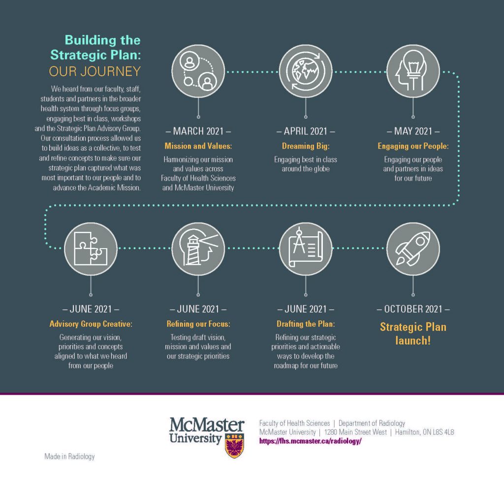
Our Strategic Plan
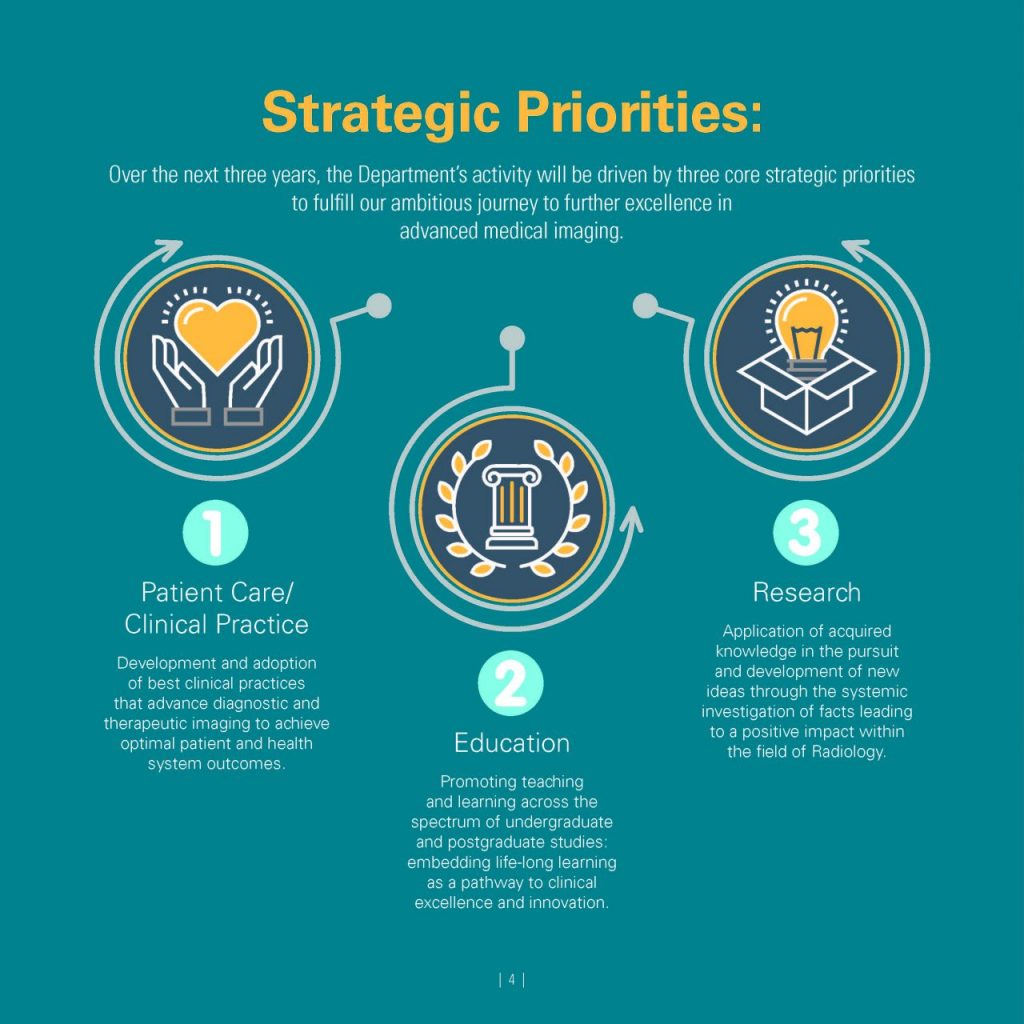
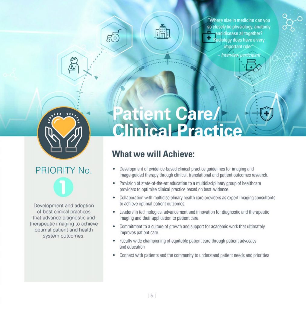
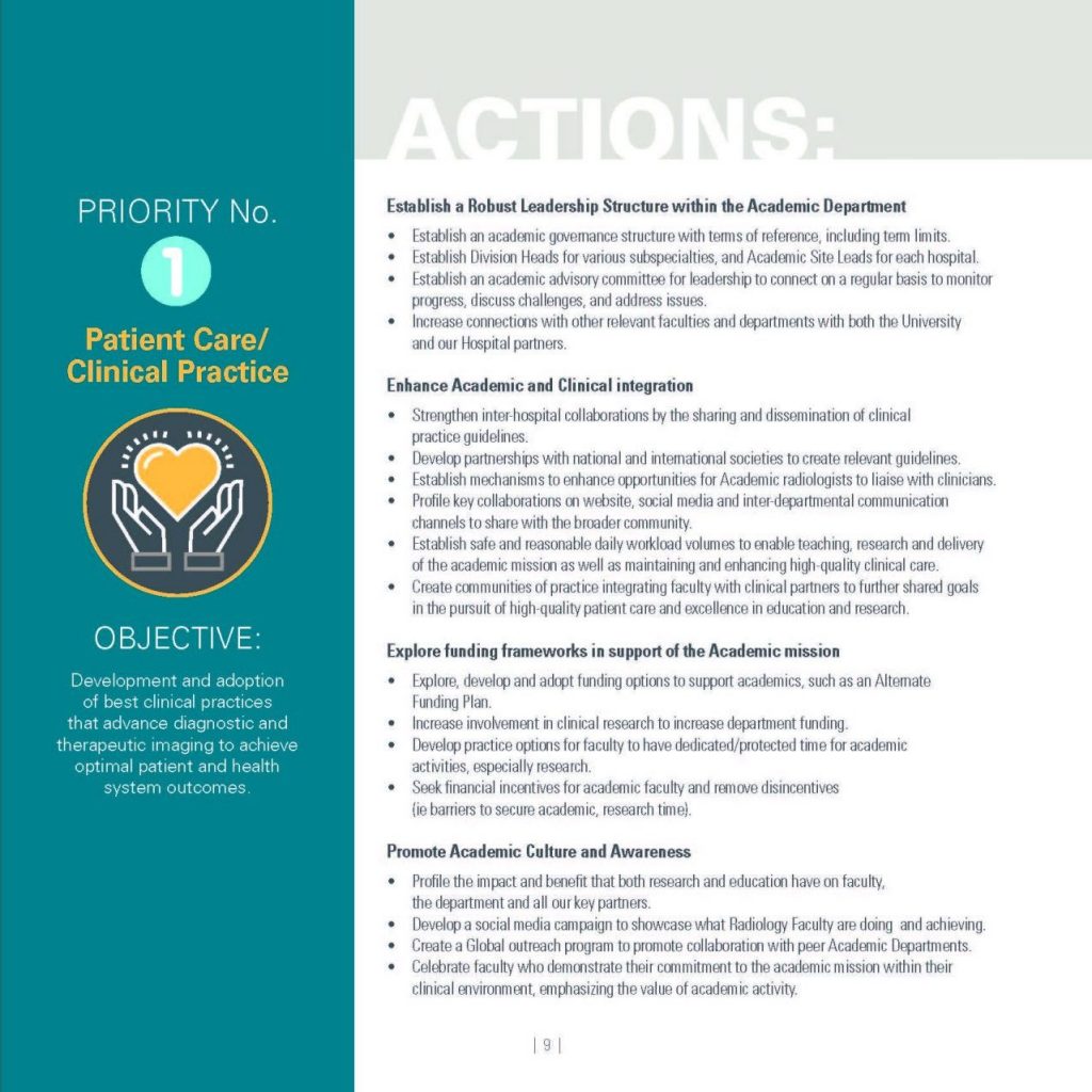
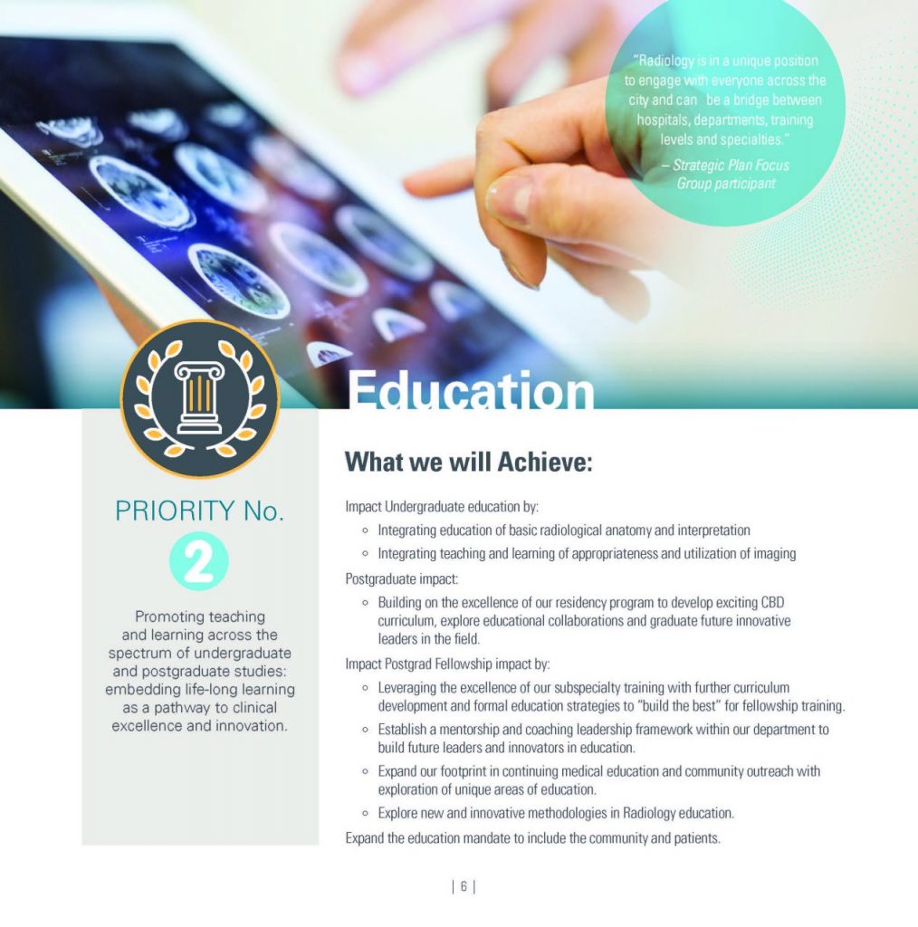
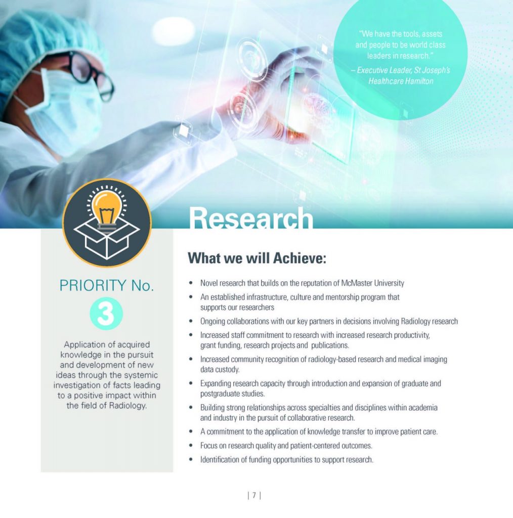
The Academic Department of Radiology 2022-25 Strategic Plan: Knowledge Advancing Healthcare represents the collective voice of our faculty, students, staff, and community partners who contributed meaningful input to this inspirational plan.
Anchored in our departmental values, we commit to a vision of advancing excellence in clinical care, research, and education to become global leaders in Radiology.
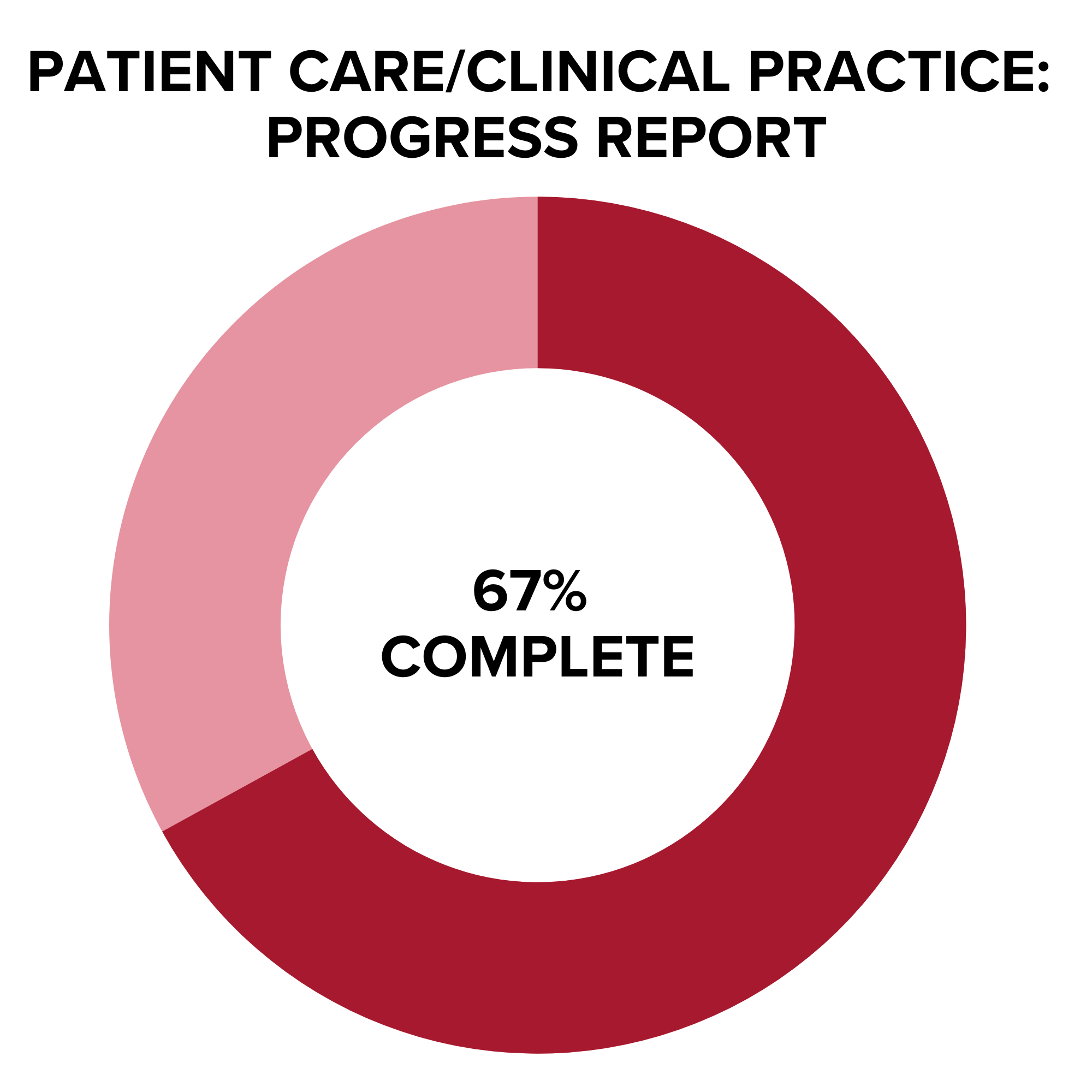
You are invited to read the plan itself and learn more about the innovative ways we will propel medical imaging to inspirational levels over the next three years. Our faculty, staff and system partners are now working to implement the transformational commitments of this plan. Updates on our progress will be communicated regularly through our departmental newsletter and posted on this site – stay tuned!
Learn more about our Strategic Plan: Department of Radiology Strategic Plan 2022 – 2025
Clinical Partners
Information Box Group
St. Joseph's Healthcare Hamilton - Learn More
St. Joseph’s Healthcare Hamilton (SJHH) provides tertiary, secondary and ambulatory health care services. SJHH is home to the multi-million dollar Father Sean O’Sullivan Research Centre, the prestigious Firestone Institute for Respiratory Health and the high-tech Brain-Body Institute. It is recognized as the regional centre of excellence for kidney and urinary diseases, respiratory medicine and thoracic surgery, as well as head and neck surgery. The hospital’s Department of Diagnostic Imaging has undergone major changes with the expansion of its programs, services and equipment, as well as redevelopment of a brand new Diagnostic Imaging department in the hospital’s new tower.
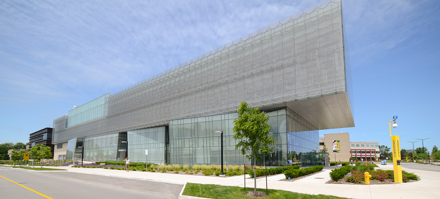
Our department of Diagnostic Imaging at St. Joseph’s Healthcare services the above programs. We have recently moved into a brand new hospital wing, shown in the picture below. Our department assets include with state of the art imaging equipment and a bright, new radiology space. We recently installed a 3.0 T clinical MR unit, in addition to our existing 1.5 T unit. We also have 2 multi-slice CTs, as well as state-of-the-art interventional suites and ultrasound equipment. We have 14 faculty members, most with fellowship specialty training. Faculty fellowship training expertise includes Musculoskeletal, Body Imaging, Cross-sectional, Interventional, Chest and Breast Imaging. Imaging rotations at this site offer a great spectrum of interesting cases.
Niagara Regional Campus - Learn More
The campus is located in a prominent city that is experiencing high economic growth from public and private investments in recreation, culture, traditional infrastructure, manufacturing, and commercial and residential development. It’s a great time for a student to be in Niagara! Find out more at: Niagara Regional Campus
Waterloo Regional Campus - Learn More
The Waterloo Regional Campus is located in the University of Waterloo Health Sciences Campus at the corner of King and Victoria Streets in the heart of the Innovation District in downtown Kitchener, Ontario.
Students at the Waterloo Regional Campus have the unique experience of living and learning in one of the fastest growing regions in the country. Find out more at: Waterloo Regional Campus
Our Committees
Information Box Group
Tenure & Promotion Committee - Learn More
Tenure & Promotion Committee – Terms of Reference
Members:
- Dr. Julian Dobranowski
- Dr. Karen Finlay
- Dr. Hema Choudur
- Dr. Stefanie Lee
- Dr. John O’Neill
- Dr. Ali Yikilmaz
Undergraduate Radiology Curriculum Committee (URCC) - Learn More
Undergraduate Radiology Curriculum Committee (URCC) – Terms of Reference
Members:
- Chair: Dr. Victoria Tan
- MF1 coordinator: Dr. Victoria Tan
- MF3 coordinator: Dr. Mostafa Alabousi
- MF4 Neuro coordinator: Dr. Zonia Ghumman
- MF4 MSK coordinator: Dr. Adam Legge
- IF coordinator: Dr. Prasa Gopee-Ramanan
- Clerkship Director: Dr. Patrick Kennedy
- Elective Director: Dr. Oleg Mironov
- Research Director: Dr. Stefanie Lee
- Resident Representative: Dr. Brian Wang
- Resident Representative: Dr. Niharika Shahi
- Resident Representative: Dr. Scott Caterine
- Resident Representative: Dr. Lucy Samoilov
- Resident Representative: Dr. Sami Adham
- Resident Representative: Dr. Vineeth Bhogadi
- Resident Representative: Dr. Alex Pozdnyakov
- Resident Representative: Dr. Katherine Li
- Resident Representative: Dr. Arbaaz Patel
- Medical School Representative: Nikhil Patil
- Medical School Representative: Benjamin Katzman
- Medical School Representative: Levi Burns
- Medical School Representative: Rod Parsa
- Medical School Representative Joseph Steinman
Radiology Post Graduate Committee (RPC) - Learn More
Radiology Post Graduate Committee (RPC) – Terms of Reference
Members:
- Dr. Yoan Kagoma – Program Director
- Dr. Natasha Larocque – APD, CBME Lead
- Dr. Ali Yikilmaz – MUMC site rep
- Dr. Milita Ramonas – JH site rep
- Dr. Tom Mammen – HGH site rep
- Dr. Euan Stubbs – SJH site rep
- Chief residents (Dr. Brian Wang, Dr. Scott Caterine)
- Jr. Resident Reps (Dr. Jason Yao, Dr. Vineeth Bhogadi)
- Cheney Matteliano – Program Assistant
Research Advisory Committee (RAC) - Learn More
Research Advisory Committee (RAC)
Members:
- Dr. Michael Noseworthy (Co-Chair)
- Dr. Katherine Zukotynski (Co-Chair)
- Dr. Nina Stein
- Dr. David Koff
- Dr. Christian van der Pol
- Dr. Mashael Alhrbi
- Dr. Steffan Frosi Stella
- Dr. Oleg Mironov
- Kelly van Staalduinen (On Leave)
- Gursimran Singh
Our Authors
 Editors: Michael Patlas
Radiologists in emergency department settings are uniquely positioned to identify and provide effective, appropriate care to vulnerable patient populations. Emergency Imaging of At-Risk Patients fills a void in the literature by illustrating challenges in emergency and trauma imaging of vulnerable patients using a head-to-toe approach. Drawing on the vast clinical experience of emergency and trauma radiologists from the largest academic medical centers across North America, this reference presents basic and advanced emergency imaging concepts, relevant case studies, current controversies and protocols, and subtle imaging findings that help guide clinicians to efficient and accurate diagnoses and treatments.
Link: Emergency Imaging of At-Risk Patients: General Principles
 Editors: Michael Patlas, Douglas S. Katz, Mariano Scaglione
This superbly illustrated book describes a comprehensive and modern approach to the imaging of abdominal and pelvic emergencies of traumatic and non-traumatic origin. The aim is to equip the reader with a full understanding of the roles of advanced cross-sectional imaging modalities, including dual-energy computed tomography (DECT) and magnetic resonance imaging (MRI). To this end, recent literature on the subject is reviewed, and current controversies in acute abdominal and pelvic imaging are discussed. Potential imaging and related pitfalls are highlighted and up-to-date information provided on differential diagnosis. The first two chapters explain an evidence-based approach to the evaluation of patients and present dose reduction strategies for multidetector CT imaging (MDCT). The remaining chapters describe specific applications of MDCT, DECT, and MRI for the imaging of both common and less common acute abdominal and pelvic conditions, including disorders in the pediatric population and pregnant patients. The book will be of value to emergency and abdominal radiologists, general radiologists, emergency department physicians and related personnel, general and trauma surgeons, and trainees in all these specialties.
Link: MDCT and MR Imaging of Acute Abdomen

Editors: Michael N. Patlas, Douglas S. Katz, Mariano Scaglione
This book describes and illustrates the gamut of errors that may arise during the performance and interpretation of imaging of both nontraumatic and traumatic emergencies, using a head-to-toe approach. The coverage encompasses mistakes related to suboptimal imaging protocols, failure to review a portion of the examination, satisfaction of search error, and misinterpretation of imaging findings. The book opens with an overview of an evidence-based approach to errors in imaging interpretation in patients in the emergency setting. Subsequent chapters describe errors in radiographic, US, multidetector CT, dual-energy CT, and MR imaging of common as well as less common acute conditions, including disorders in the pediatric population, and the unique mistakes in the imaging evaluation of pregnant patients. The book is written by a group of leading North American and European Emergency and Trauma Radiology experts. It will be of value to emergency and general radiologists, to emergency department physicians and related personnel, to general and trauma surgeons, and to trainees in all of these specialties.
Link: Errors in Emergency and Trauma Radiology
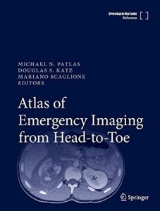
Editors: Michael N. Patlas, Douglas S. Katz, Mariano Scaglione
This reference work provides a comprehensive and modern approach to the imaging of numerous non-traumatic and traumatic emergency conditions affecting the human body. It reviews the latest imaging techniques, related clinical literature, and appropriateness criteria/guidelines, while also discussing current controversies in the imaging of acutely ill patients.
The first chapters outline an evidence-based approach to imaging interpretation for patients with acute non-traumatic and traumatic conditions, explain the role of Artificial Intelligence in emergency radiology, and offer guidance on when to consult an interventional radiologist in vascular as well as non-vascular emergencies. The next chapters describe specific applications of Ultrasound, Magnetic Resonance Imaging, radiography, Multi-Detector Computed Tomography (MDCT), and Dual-Energy Computed Tomography for the imaging of common and less common acute brain, spine, thoracic, abdominal, pelvic and musculoskeletal conditions, including the unique challenges of imaging pregnant, bariatric and pediatric patients.
Written by a group of leading North American and European Emergency and Trauma Radiology experts, this book will be of value to emergency and general radiologists, to emergency department physicians and related personnel, to obstetricians and gynecologists, to general and trauma surgeons, as well as trainees in all of these specialties.
Link: Atlas of Emergency Imaging from Head-to-Toe

Editors: Michael N. Patlas, Douglas S. Katz, Mariano Scaglione
This book presents a comprehensive and modern approach to the imaging of nontraumatic and traumatic emergencies in pregnant patients. Readers will find a careful review of the relevant imaging-related clinical literature, explanation of imaging appropriateness criteria and guidelines, and enlightening discussion of current controversies in the emergency imaging of obstetric patients. The opening chapter discusses general principles of emergency imaging during pregnancy and offers an overview of an evidence-based approach to imaging interpretation. The remainder of the book describes specific applications of ultrasound, MRI, radiography, and MDCT for the imaging of common as well as less common acute brain, spine, thoracic, abdominal, and pelvic conditions during pregnancy. Clear guidance is offered on the unique challenges that may be encountered during such imaging. Emergency Imaging of Pregnant Patients is written by a group of leading North American and European emergency and trauma radiology experts. It will be of value to emergency and general radiologists, to emergency department physicians and related personnel, to obstetricians and gynecologists, to general and trauma surgeons, and to trainees in all of these specialties.
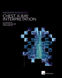 Editors: Julian Dobranowski, Alexander J. Dobranowski, Anthony J. Levinson
Divided into 3 sections. The first section provides a description of the technology, the patient journey, how the x-ray image is created, and the elements needed for interpretation. The second section analyses how normal anatomical structures appear on an x-ray and how pathology modifies that appearance. The third section concentrates on the interpretive process and specific problems related to chest imaging. Throughout the book, the authors incorporate evidence-based techniques to enhance learning and skill development. The use best practices in designing graphics for learning when creating the instruction-al images. The sections of the book are deliberately segmented and sequenced to facilitate transfer of concepts and skills. Each chapter has a structured introduction as an advanced cognitive organizer to help situate the material, as well as a case-based example to activate prior knowledge, provide authentic context, and reinforce the material. To improve application of the knowledge in the clinical setting, the book is filled with valuable checklists and job aides that provide performance support tools.
2in1 Discover Radiology: Chest X-Ray Interpretation BOOK + MOBILE APP is the first in a series of radiology textbooks concentrating on the teaching of x-ray interpretation through a focus on anatomy, radiological anatomy, concepts of pathology, differential diagnosis and important concepts and competencies.
Link: Discover Radiology: Chest X-Ray Interpretation
Polish Cover:

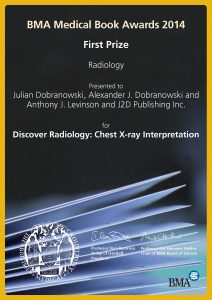
Awards:

 Editors: Julian Dobranowski, David A. Stringer, Sat Somers, Giles W. Stevenson, William M. Thompson
Dr. Dobranowski and his associates are to be highly commended for this excellent manual. I am not aware of a similar text covering the subject. Although all of us perform gastrointestinal studies in a differ ent manner, this text provides an excellent overview. The reader will discover that the text is especially well written and focuses on the important issues relating to GI contrast studies. Because Dr. Steven son’s group performs endoscopic procedures, they are included in the manual. These authors are recognized scholars and leaders in gastrointesti nal radiology. Thus, it is easy to understand why the manual is so well done. I am particularly impressed with the emphasis placed on the patient-radiologist relationship before, during, and after completion of a study. All of us who teach gastrointestinal radiology are concerned about the decline in the number of gastrointestinal contrast studies. We are not sure how we can continue to teach our residents the proper tech niques and maintain high-quality teaching programs in gastrointesti nal radiology. A manual of this type is thus timely and appropriate. The manual will be a valuable addition to the library of all radiologists. It will be particularly useful for residents who are learning how to per form GI contrast studies.
Link: Procedures in Gastrointestinal Radiology
 Editor: John M. D. O’Neill
Proper ultrasound examination and interpretation hinges on thorough knowledge of the relevant anatomy, artifacts, and technique. This book provides an excellent foundation by going beyond pathology and concentrating on these fundamentals. Basic physics and artifact recognition and prevention are outlined. Chapters review essential anatomy and include images and tables that highlight relevant bones, ligaments, tendons, muscles, and nerves. Sites of attachment and the best positions for examination are also noted. Technique is presented via a three-tiered approach and photographs of patients in the transducer position are matched with the resulting ultrasound images and complementary anatomical overlays.

Editor: John O’Neill
This book offers an excellent review of the various rheumatological conditions, both common and uncommon, that may present on imaging on a daily basis. The book uses a unique format that will be beneficial for clinicians, radiologists, medical students, and consultant staff. The text is written by both rheumatology and radiology staff to provide a balanced approach. A clinical overview and the common clinical presentations are briefly reviewed for each condition followed by a more detailed discussion of imaging findings produced by the various imaging modalities, including radiographs, ultrasound, MRI, CT, and nuclear medicine. This book details the imaging of normal musculoskeletal anatomy and pathology; discusses image-guided musculoskeletal interventions; and examines disorders such as rheumatoid arthritis, connective tissue disease, osteoarthritis, osteonecrosis, infection-related arthritis, soft tissue calcification, and bone and synovial tumors. Featuring over 600 multi-part, high-resolution images of rheumatic diseases across current imaging modalities, Essential Imaging in Rheumatology offers up-to-date and complete information on the imaging of these disorders.
Link: Essential Imaging in Rheumatology

Editors: J O’Neill & Walter Maksymowych
A practical comprehensive visual guide to the complex imaging appearance of axial spondyloarthritis in clinical practice.
This atlas is a collaboration between diagnostic imaging and rheumatology, providing an extensive array of images that illustrate the diverse pathology that occurs in the SIJ and spine in patients with axial SpA. While the primary focus is on MRI there are also examples of plain radiographic and CT findings to highlight the comparative advantages and disadvantages of different imaging modalities. Additional images also illustrate the potential pitfalls of MRI and the wide imaging and clinical differential diagnosis that can be confused with axSpA.
Link:Imaging Atlas of Axial Spondyloarthritis
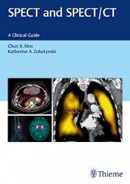
Editors: by Chun K. Kim and Katherine A. Zukotynski
Internationally renowned authorities in the field of hybrid imaging contribute firsthand expertise on the practical application of single-photon emission computed tomography (SPECT) and SPECT/CT. By combining clear anatomic markers from CT with functional knowledge from SPECT, SPECT/CT provides added value for patient evaluation and is becoming increasingly prevalent in routine clinical practice. Indeed, hybrid imaging is touted by many as a game changer in nuclear medicine.
The first two chapters of this book provide a foundation for understanding SPECT and SPECT/CT technological principles, including the associated radiopharmaceuticals. The remaining chapters detail the utility of SPECT and SPECT/CT in clinical practice including neuroscience and pediatrics, as well as specific pathologies. The book concludes with in-depth discussion of select case studies.
 Editor: Sriharsha Athreya
This book is a concise introduction to the interventional radiology field and is designed to help medical students and residents understand the fundamental concepts related to image-guided interventional procedures and determine the appropriate use of imaging modalities in the treatment of various disorders. It covers the history of interventional radiology; radiation safety; equipment; medications; and techniques such as biopsy and drainage, vascular access, embolization, and tumor ablation. The book also describes the indications, patient preparation, post-procedure care, and complications for the most common interventional radiology procedures.
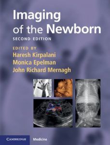 Editors: Haresh Kirpalani, Monica Epelman and John Richard Mernagh
This fully revised new edition of a popular practical guide provides a concise introduction to radiology in neonates, covering the full range of problems likely to be encountered in the neonatal ICU. The material is presented in atlas format, with concise text descriptions to provide a quick overview of the indications, utility, appearances and interpretation of images of common neonatal pathology. Numerous high-quality images enable easy ‘matching’ with clinical cases faced by the reader. New to this edition: • Images updated throughout to reflect improvements in equipment and scanning techniques • Expanded chapters on cardiovascular problems, bone and prenatal ultrasound • New chapters on clinical utility of procedures, metabolic and inborn errors of metabolism, and antenatal diagnosis of common abnormalities Concise and practical, this is an essential training resource for all those who work in the neonatal ICU, including pediatric residents and trainees, junior radiologists and nurse practitioners.
Link: Imaging of the Newborn
 Editors: Haresh Kirpalani, Monica Epelman and John Richard Mernagh
Diagnostyka obrazowa nowarodka (published in Polish), Medipage, 2015
 Editors: Haresh Kirpalani, Gerald Gill, and John Mernagh
A practical handbook to the significant features seen on images of common neonatal pathology. Over 230 superb images illustrate each condition while radiological features are identified together with clinical features, indications, and contraindications for further imaging. This handy guide identifies radiological features within their clinical context.
Link: Imaging of The Newborn Baby
 Editors: Eli T. Tshibwabwa, Glen Oomen, John Mernagh, Craig Coblentz and Bruce Wainman
Ultrasound Anatomy – An Imaging Resource for Medical Students in their Journey through the Medical Foundations
Educational Program in Anatomy, McMaster University Faculty of Health Sciences. 2008.
 Editor: John Richard Mernagh
ACR Teaching File in Obstetrical Ultrasound American College of Radiology, 2008.
Our Leadership
The Academic Department of Medical Imaging has been privileged to build world-class educational programming for our medical students, residents, and fellows, contribute to excellence in research that has global impact, and pride themselves on delivering high-quality patient care to the people in our community every day. A large portion of this excellence and forward momentum is credited to our faculty leadership.
Our innovative leadership team consists of dedicated, focused, and talented faculty. Working with our team, students and system partners, our leaders come together to make a difference locally, provincially, nationally and around the globe.
Meet our Faculty Leadership Team:
Information Box Group

Chair
Dr. Julian Dobranowski

Associate Chair Education
Dr. Karen Finlay

Undergraduate Director
Dr. Victoria Tan

Residency Director
Dr. Yoan Kagoma

Neuroradiology Residency Director
Dr. Crystal Fong

Fellowship Director
Dr. Ehsan Haider

Research Co-Chair
Dr. Michael Noseworthy

Research Co-Chair
Dr. Katherine Zukotynski
Our Resources
Resource Listing
POP-UP
Reports
POP-UP
Regional Attractions
Regional Attractions
- Canadian Medical Association (CMA)
- Canadian Medical Protective Association (CMPA)
- Canadian Resident Matching Service (CaRMS)
- College of Physicians and Surgeons of Ontario (CPSO)
- Ontario Medical Association (OMA)
- Professional Association of Internes and Residents of Ontario (PAIRO)
- Royal College of Physicians and Surgeons of Canada (RCPSC)
- The City of Hamilton
- The Province of Ontario
- The Government of Canada
- Algonquin Park (Ontario’s best known Provincial Park)
- National Gallery of Canada
- RadList.com
- Royal Botanical Gardens (RBG)
- Royal Ontario Museum (ROM)
- Greater Toronto Airport Authority
- Canada 411 (Phone Directory)
- Postal Code Lookup by Canada Post Corporation
- Weather Conditions & Forecasts by Environment Canada
Affiliated Health Care Centers in Hamilton
Selected Radiology Journals and Sites
WEBSITE
Medportal
POP-UP
Useful Radiology Weblinks
Useful Radiology Weblinks
- MIIRCAM
- Canadian Association of Radiologists (CAR)
- American College of Radiology (ACR)
- American Roentgen Ray Society (ARRS)
- Armed Forces Institute of Pathology (AFIP)
- Aunt Minnie (Radiology News and Teaching)
- Canadian Heads of Academic Radiology for Radiology in Canadian Universities
- Eurorad (Teaching Cases)
- Neuroanatomy Lab (Interactive Neuroanatomy)
- Radiological Society of North America (RSNA)
- Society of Chairmen In Academic Radiology Departments Fellowship Match
- Society of Nuclear Medicine (SNM)
- Society of Thoracic Radiology (STR)
- Uniformed Services University of the Health Sciences (USUHS)
- University of Alberta
- University of British Columbia
- University of Iowa Physiological Imaging
- University of Iowa Department of Radiology
- University of Michigan
- University of Toronto
- University of Washington


